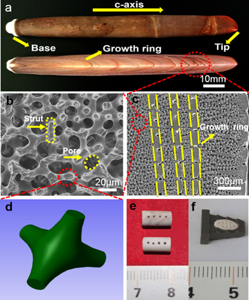
Figure 2. Section view of original sea urchin spines (H. mammillatus)(a), SEM micrographs of inner structures of sea urchin spines (b−e), and configuration of a truncated conical shell for a strut in the sea urchin spine (f) and machined sea urchin spine samples as bone implants (g and h).
| 